


STROKE and QEEG
Stroke refers to all the lesions of brain tissue caused by abnormalities in blood vessels, located in the brain. This
can be classified into cerebral infarction, which is due to cerebrovascular blockage, and cerebral hemorrhage,
caused by the burst of brain blood vessels.
According to previous QEEG studies, it has been reported that an abnormal increase of slow-waves is observed in
the area where the brain injury occurred in stroke. This is observed according to the degree of ischemic injury as
below.
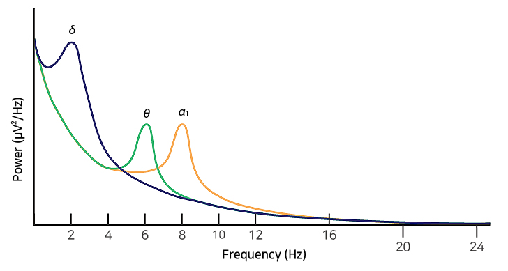
Very slow delta waves in severe area
Slow theta waves in minor areas
Slow alpha waves in the surrounding areas
Using QEEG for strokes can range the recovery of brain function with differentiation and treatment according to
the damaged area. This can increase treatment success rate through monitoring before and after treatment.
CASE REVIEW
Participant information
– Age : 55 / Male / Diagnosed with stroke(Cerebral infarction)
– Cognitive function decreased after three months of the onset of left middle cerebral artery infarction.
| Band Power – Topomap
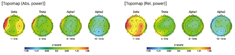
Compared to healthy people, a decrease of delta wave was shown in the left frontal and temporal lobes,
which are the site of the lesions.
| Power Spectrum
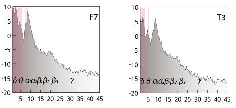
| Component
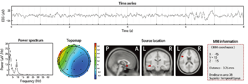
Compared to healthy people, in the power spectrum and component, a delta wave was commonly
observed in the left frontal and temporal lobes, which are the site of the lesions.
| 3D VIEW
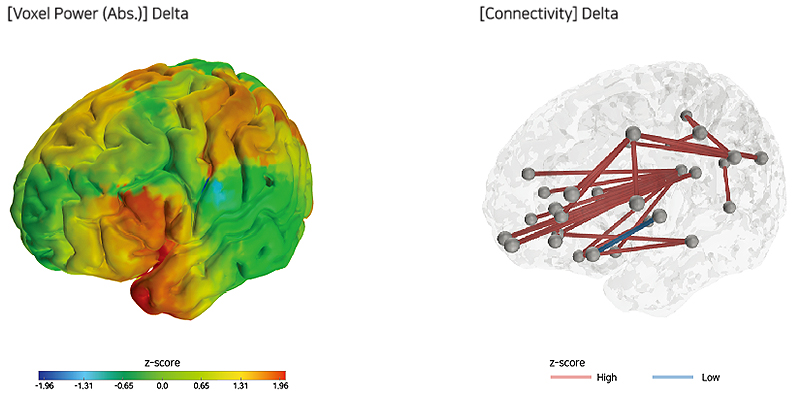
Compared to healthy people, an increase of delta wave and Connectivity was shown in the left frontal and
temporal lobes, which are the site of the lesions.


